Vascular access
Vascular access
Fistula, graft, and catheter planning, with daily checks for dysfunction and infection.
A reliable vascular access is essential for people with end-stage kidney disease. The source estimates that access problems account for approximately one quarter of hospital admissions among haemodialysis patients and are a major cause of morbidity.
The modern history of long-term dialysis access began in 1960, when Clyde Shields became the first person with chronic kidney failure to receive maintenance haemodialysis through a shunt developed by Dr Belding H. Scribner, who was born in Chicago on 18 January 1921 and died on 19 June 2003.
Scribner’s 1960 design placed Teflon and silicone-rubber cannulae in the patient’s forearm and connected them externally. For the first time, repeated haemodialysis could continue over many months. Shields lived with chronic kidney failure for another eleven years and died of heart disease in 1971.
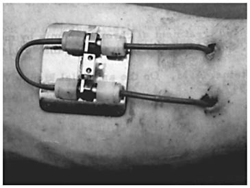
This revolutionary invention made long-term treatment of end-stage kidney failure possible for the first time.
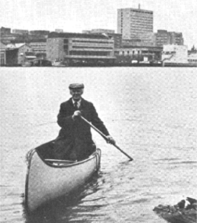
Scribner died at the age of 82 after drowning in the lake he crossed by canoe on his way to work.
Dialysis units need strategies that reduce complications related to vascular access, including:
Favouring an arteriovenous fistula (AVF) over a central venous catheter when it is appropriate for the individual patient.
Planning access early in people with progressive chronic kidney disease.
Reducing infection and thrombosis.
Regular clinical monitoring to detect an access at risk of stenosis, thrombosis, or failure.
Synthetic arteriovenous grafts have higher rates of thrombosis and infection and require more surgical or endovascular interventions than mature native fistulas. They should therefore be used selectively. The source reports better long-term patency for primary AVFs across age groups and a substantially lower infection burden than with grafts.
When to plan a fistula or other vascular access
Early referral to kidney care and timely access planning reduce the complications of emergency surgery and urgent catheter placement when haemodialysis becomes necessary.
A native fistula needs time to mature before it can be used. The source recommends beginning preparation roughly a year before dialysis is expected and creating the AVF at least 6–12 months beforehand. A synthetic graft can often be placed about one month before anticipated use. Tunnelled and temporary catheters are best inserted only when needed because complications—especially infection—rise with catheter exposure. Modern planning should be individualised rather than based on one eGFR threshold or a universal “fistula first” rule.
Clinical assessment before access creation
The assessment includes:
A complete cardiovascular examination, including pulses and bruits or murmurs over the heart and limbs.
Comparison of blood pressure and pulses between the limbs.
An Allen test to assess ulnar-artery supply: compress both the radial and ulnar arteries at the wrist until the hand becomes pale, then release only the ulnar artery and observe reperfusion of the whole hand. Failure of colour to return suggests inadequate ulnar patency or flow.
Assessment for oedema.
Central venous stenosis is not uncommon after previous central venous catheterisation. It is therefore important to look for this complication before access surgery.
Some units use duplex ultrasound or angiographic imaging before surgery in patients with oedema, previous surgery, or suspected vascular disease; some use routine vessel mapping. Clinical and ultrasound vein mapping can document vessel location and diameter from wrist to axilla and help identify the best vein for anastomosis. The source gives a desired vein diameter above 2.5 mm and arterial diameter above 2 mm, without stenosis.
People with chronic kidney disease and the staff caring for them should protect potential access veins. Forearm veins should be avoided for routine intravenous cannulation or venepuncture whenever possible, especially in the arm intended for a fistula. Subclavian venous catheterisation should be avoided in patients with kidney disease because it can compromise future access through central venous stenosis.
Permanent vascular access
Permanent access can be created as a native AV fistula or with a synthetic or biological graft. An AVF is a subcutaneous anastomosis between an artery and a vein, usually in the forearm. Over several months, the vein dilates and its wall thickens—a process called arterialisation.
An arteriovenous graft is an artificial connection between an artery and a vein. It is commonly made from polytetrafluoroethylene (PTFE, Teflon®), although biological materials such as bovine vessels can also be used.
A tunnelled central venous catheter, such as a Demers-type catheter, can provide longer-term access when vessels are too small, fistula creation has failed, or no surgical access is feasible. It is generally not preferred when a suitable fistula or graft is possible because catheter-related complications are more frequent.
Arteriovenous fistula
For a suitable patient, a mature native fistula generally offers better long-term patency and fewer complications than a graft or long-term catheter. Unlike a graft, its flow and usability may continue to improve after maturation.
An AVF is usually created as distally as feasible in order to preserve proximal vessels and reduce complications associated with upper-arm access. Options progress from distal to proximal:
Radiocephalic fistula at the wrist—the classic Brescia–Cimino fistula introduced in the 1960s and usually the first option when anatomy is suitable.
Radiobasilic fistula at the wrist or distal forearm.
Brachiocephalic fistula at the elbow or upper arm.
Brachiobasilic fistula, which may require a second operation to transpose or superficialise the deeply situated vein.
If upper-limb native options are unsuitable: forearm grafts, femoral fistulas or grafts, and axillo-axillary grafts.
After a bridge graft fails, a native fistula may occasionally become possible because proximal veins have enlarged. AVFs need to be created far enough in advance—at least 3–4 months in the source—to permit maturation and correction of problems. Published cohorts have reported failure to mature in up to 40% of fistulas, although rates differ substantially by population and definition.
Synthetic arteriovenous grafts
A graft is used when creation of a usable native fistula is not feasible. The artery and vein are joined by a straight or looped tube, usually made of PTFE. Its porous structure allows fibroblast ingrowth and incorporation into the subcutaneous tissue.
Other graft materials include transposed natural veins, human umbilical vein, bovine mesenteric vein products such as ProCol®, and other synthetic or biological conduits.
A common configuration is a loop roughly 10 cm long between the brachial artery and a cephalic or other suitable vein in the forearm or upper arm.
A straight forearm graft may also connect the radial artery and cephalic vein.
A short graft can deteriorate when a limited area is punctured repeatedly.
An excessively long graft may have higher resistance, lower flow, and a greater risk of thrombosis.
Grafts are generally easier to cannulate than fistulas and can often be used relatively soon after surgery—commonly after about 14 days. Earlier cannulation may be possible for selected early-cannulation grafts, but with a standard graft it can increase bleeding and perigraft haematoma, which may itself promote thrombosis.
As noted above, a graft should not automatically be the first option.
Access blood flow
The source gives an average blood flow of 800–1,200 mL/min through a forearm graft.
Fistula flow is more variable; the source states that it should generally exceed 600 mL/min. Flow thresholds must be interpreted with the access type, trend, clinical examination, and dialysis performance.
Disadvantages of permanent vascular access
Disadvantages of an AV fistula
Slow maturation and a long interval before reliable cannulation.
Failure to mature: the vein remains small and cannot be cannulated because flow is inadequate.
Cannulation can be more difficult than with a graft.
The access may enlarge considerably over time.
Aneurysm formation.
Cosmetic concerns.
Dialysis access steal syndrome and distal ischaemia.
Disadvantages of a graft
Surgery is more complex than for a straightforward native fistula.
Substantially higher infection risk.
The source reports an approximately sixfold higher thrombosis risk than for an AVF.
Stenosis at an anastomosis, especially the venous anastomosis.
A limited expected lifespan, cited as approximately 3–5 years.
Removal can be difficult.
Skin erosion. Once the skin over a graft erodes, the graft commonly becomes infected and rarely heals with antibiotics alone. A new access at another site and removal of the exposed segment may be required, allowing the wound to heal by secondary intention.
Creation of permanent vascular access
Creation of a fistula
Radial access: the radial artery is joined to the cephalic vein at the wrist, usually end-to-side; a side-to-side anastomosis may be used when the vessels overlap and lie close together.
Brachial access: use of the brachial artery produces higher flow and therefore a greater risk of steal and oedema.
Oedema is common soon after surgery, particularly with a side-to-side anastomosis, because venous pressure rises in the distal vein beyond the anastomosis. This is less prominent with an end-to-side venous connection.
The cephalic vein can be mobilised and joined to the brachial artery. Another option is the deep communicating vein arising from the median antecubital vein.
Dehydration and hypovolaemia should be avoided before surgery. Patients may need adequate intravenous fluid and a normal to slightly high blood pressure; both marked hypotension and severe hypertension are undesirable.
The source suggests allowing a dialysis patient to remain slightly above dry weight before access surgery.
Local or regional anaesthesia is usually used for distal access surgery.
After surgery, hypotension should be avoided. The source suggests withholding selected antihypertensive medication, delaying haemodialysis for at least 24 hours when feasible, and temporarily increasing target weight during the following week if needed to reduce early fistula thrombosis. These decisions require individual clinical judgement.
Evidence has not established that routine aspirin, another antiplatelet agent, or systemic anticoagulation improves long-term access survival after surgery.
Measures that may support fistula maturation
Hand and arm exercises may support maturation or at least improve the patient’s awareness of the access.
Avoid additional exertion when a postoperative haematoma or oedema is present until swelling subsides.
If there is concern that the new fistula has been injured, temporary single-needle dialysis and avoidance of repeated puncture may be prudent.
Creation of a bridge graft
After suitable vessels have been exposed, the graft is tunnelled under the skin and anastomosed to the artery and vein. Twisting and kinking must be avoided.
Systemic anticoagulation is rarely required. Evidence is conflicting for aspirin or low-dose warfarin as prophylaxis against graft thrombosis. Careful maintenance of euvolaemia and avoidance of hypotension before and after surgery are more important.
Stenosis of fistulas and grafts
Venous stenosis
Venous stenosis is less common in an AVF than in a graft but can be more severe. It usually occurs near the anastomosis, although it may be more remote or lie anywhere along the draining vein. Intimal and fibrous hyperplasia are major mechanisms. Stenoses can also develop within grafts.
Possible clues include rising venous pressure, which is not very sensitive; falling access flow measured by Doppler; and recirculation, which tends to be a later but more specific sign.
An intragraft stenosis may not produce the same pressure change or recirculation pattern as an outflow stenosis.
There is no evidence that correcting a stenosis without haemodynamic significance—described here as less than 50% diameter reduction—improves outcomes or lowers thrombosis rates.
Repair while the access remains patent is generally more successful than intervention after thrombosis. The historical figures quoted in the source are only 50% graft patency four weeks after angioplasty of a thrombosed graft, compared with 80% patency at 28 weeks after angioplasty of a non-thrombosed access.
The source states that surgical revision or angioplasty may be less effective in some AVFs than in grafts, depending on lesion anatomy.
Angioplasty can be difficult in a longstanding graft with pronounced neointimal hyperplasia.
Stents were used more freely in the past. Subsequent fibrosis can make later thrombectomy difficult and may leave recurrent stenosis less amenable to surgical correction. Modern stent-graft use is lesion-specific.
Thrombosis of fistulas and grafts
Thrombosis is reported roughly six times more often in grafts than in AVFs, although loss of a fistula may have greater long-term consequences.
It is usually secondary to a stenosis or low inflow from the feeding artery.
Thrombus can be removed surgically with a balloon-tipped Fogarty embolectomy catheter, but the underlying structural cause—identifiable in approximately 80%–90% of cases according to the source—must also be treated.
Endovascular thrombectomy and angioplasty are often preferred for thrombosed access with a correctable stenosis.
Local thrombolysis with tissue plasminogen activator, alteplase, or historically streptokinase can also be used, often with heparin. The source describes tPA 0.5–2 mg followed by 0.5 mg increments, delivered through a standard or pulse-spray catheter over several hours. These are specialist procedural doses, not instructions for unsupervised use.
Success falls as the thrombus ages, particularly after about 48 hours.
Antiplatelet treatment such as clopidogrel or aspirin with dipyridamole, and anticoagulation with a vitamin K antagonist such as warfarin, have not consistently reduced access thrombosis and can increase bleeding.
Small trials have suggested that omega-3 polyunsaturated fatty acids may reduce graft-thrombosis rates.
Monitoring for stenosis or thrombosis
The source estimates that approximately 20% of patients with an AVF require an access intervention each year, with still higher rates for grafts. Access failure is a frequent cause of hospital admission.
Monitoring can include:
Access blood flow, Qa.
Static venous pressure.
Dynamic venous pressure.
Measurement of access recirculation.
A fall in delivered dialysis dose or efficiency.
A more negative arterial pressure.
Duplex Doppler ultrasound.
Clinical signs of arterial inflow stenosis include a poorly filled access, excessive collapse of the outflow vein when the arm is raised, low pump flow or repeated “sucking down” of the arterial needle segment, and an abnormal bruit along the access. Venous outflow stenosis can cause arm swelling and an abnormal high-pitched bruit or thrill along the draining vein.
One study cited in the source found that combining physical examination with routine Doppler flow measurement detected more than 80% of AVF stenoses.
Access flow rates
The source gives typical AVF flow of 500–800 mL/min and graft flow of 600–1,000 mL/min, occasionally as high as 3 L/min. A readily palpable thrill usually indicates flow above approximately 400 mL/min.
For grafts, the source describes a greater risk of thrombosis over the following six months when flow falls below 600 mL/min, or falls by more than 25% over four months and is below 1,000 mL/min.
Repeated measurements and a downward trend are more informative than a single value. The trend is often the most important predictive signal.
Flow is commonly measured in the brachial artery, where measurement is reproducible and should approximate total access flow.
Dynamic venous-pressure monitoring
A random venous-pressure value displayed by the dialysis machine is a poor marker of venous stenosis. Measurements are more useful under standardised conditions at comparable times—for example, during the first five minutes of every session at a blood flow of 200 mL/min, using needles and tubing of the same length and gauge.
Patients with pressure rising over successive sessions need access assessment or venography, as do those exceeding a locally defined threshold. Historical device-specific thresholds quoted in the source are dynamic venous pressure above 125 mmHg at Qb 200 mL/min with a 15-gauge needle on a Cobe Centrysystem® 3, and 150 mmHg with a 15-gauge needle on a Gambro AK10®.
A rising trend over consecutive sessions is more important than one isolated high value.
Measurement of recirculation
Recirculation is a relatively late predictive sign and often appears only after access flow has fallen to approximately 350–500 mL/min.
Values above 15% are considered clinically important in the source and may indicate significant stenosis. Several measurement techniques exist; false-negative results can occur, so testing must be standardised.
A fall in URR or Kt/V
Many factors affect the delivered dialysis dose, so a fall in URR or Kt/V is not specific for access stenosis.
Venography or arteriography
Definitive imaging is required when there is strong suspicion of a venous or arterial stenosis.
Venography may not display the arterial anastomosis well and can miss a lesion there. Arteriography can show the entire access more completely, including arterial inflow and venous drainage.
Other complications of fistulas and grafts
Low flow
Adequate access flow is essential for effective haemodialysis.
Low flow is also an independent risk factor for access thrombosis. It commonly requires vascular imaging and treatment of an underlying stenosis by angioplasty or surgical revision.
Oedema
Oedema is common, especially after a side-to-side venous anastomosis, because venous pressure rises in the distal vein beyond the anastomosis. Conversion to an end-to-side configuration may help. Severe cases occasionally require closure of the AVF.
Aneurysm formation
The usual cause is repeated cannulation at one site or within a very small number of sites.
Repair is generally considered when the overlying skin becomes thin, the aneurysm becomes very large, spontaneous bleeding occurs, available cannulation sites are limited, a nerve is compressed, or cosmetic impact is substantial.
Flow disturbance within an aneurysm can increase thrombosis risk.
Pseudoaneurysm
A pseudoaneurysm is a communication between the fistula or graft lumen and a confined space in the surrounding tissue. It can compromise the overlying skin and lead to poor haemostasis and prolonged bleeding after needle removal.
Repair is required when skin integrity is threatened, rupture or nerve compression is a concern, or no other safe cannulation sites remain.
Haemodialysis access-induced distal ischaemia (HAIDI)
Ischaemia of the hand and fingers can cause permanent tissue loss, small areas of infarction, or exercise-induced symptoms. It is more common in older people, those with diabetes, and patients with several previous failed accesses.
Patients should report changes in sensation or temperature and any new weakness.
Mild distal ischaemia is relatively common and may present only as a cooler hand or numbness. It often improves over time and may not require intervention.
More severe ischaemia can require urgent treatment by closing the fistula or by a procedure such as distal revascularisation with interval ligation (DRIL). In DRIL, the artery immediately distal to the AVF is ligated and a saphenous-vein bypass carries blood from a point proximal to the ligation to a more distal artery.
The HAIDI spectrum includes dialysis access steal syndrome (DASS) and ischaemic monomelic neuropathy (IMN).
Dialysis access steal syndrome (DASS)
DASS develops when the fistula or graft offers a lower-resistance pathway than the distal arterial bed, especially when arterial inflow is already limited by atherosclerosis or stenosis. Distal blood flow falls and may reverse toward the lower-resistance access.
Brachiobasilic and brachiocephalic fistulas carry the greatest risk.
Clinical severity ranges from colour change and altered sensation to coldness, pain, and marked cyanosis at rest.
Symptoms often worsen during dialysis and may improve partly when the access is compressed.
Severe DASS can lead to digital gangrene.
Access revision may reduce flow or increase resistance; alternatives include proximalisation of arterial inflow and, when necessary, access ligation.
Ischaemic monomelic neuropathy (IMN)
IMN is a rare form within the HAIDI or steal-ischaemia spectrum but has distinctive features.
It results from focal ischaemia of peripheral nerves after the blood supply is abruptly diverted away from the nerves of the forearm and hand.
The condition is uncommon and its true incidence is unknown. It occurs almost exclusively after AV access based on the brachial artery, is reported more often in women and people with diabetes, and affects several peripheral nerves in the upper limb.
Symptoms generally begin immediately after surgery. Neurological deficits dominate, often without the usual tissue signs of distal ischaemia.
There is diffuse sensory and motor impairment in the radial, ulnar, and median nerve distributions.
Associated symptoms include pain, paraesthesia, and numbness of the hand. Motor impairment can be severe, with weak wrist extension, loss of intrinsic hand-muscle function, and weak thumb opposition. Untreated deficits can progress to a characteristic claw-hand deformity.
The hand is usually warm with preserved capillary refill, and a radial or ulnar pulse—or at least a Doppler signal—remains present. This contrasts with severe classic steal ischaemia.
Infection
Infection is mainly a problem of grafts and catheters and is less common in native fistulas. It is especially frequent with temporary central venous catheters, where Gram-positive bacteraemia predominates.
Once a graft has been colonised, complete eradication of bacteria is difficult. Prolonged antibiotic treatment and removal of part or all of the graft may be required.
An AVF infection is uncommon. The source recommends managing a deep fistula infection or associated bacteraemia with the same seriousness as endocarditis, including a prolonged course of antibiotics.
The source also describes prophylactic antibiotics for patients with grafts during dental and catheter procedures; modern prophylaxis decisions should follow current local guidance and the specific procedure.
Bleeding or extravasation injury
This usually occurs when a venous needle penetrates through a graft—more commonly—or an AVF, becomes displaced during dialysis, or bleeding continues because haemostasis after needle removal is inadequate.
Before the machine stops the blood pump, rapid limb swelling can develop. Other consequences include access thrombosis, compartment syndrome, neuropathy, severe ischaemia, and secondary infection. Surgical drainage may be required.
Superior vena cava or central venous obstruction syndrome
The usual presentation is marked swelling of one arm with dilated veins over the chest, sometimes accompanied by neck and facial swelling.
Obstruction can involve the subclavian vein—most commonly—the superior vena cava, or the internal jugular veins.
It is usually secondary to previous central venous catheterisation in these vessels and only rarely to extremely high venous return from the access itself.
Initial management may be observation in selected cases. Other options include anticoagulation when thrombosis is present, venoplasty, stenting, or open surgery. The source cites low six-month patency of only 20%–40% after venoplasty and notes that a central venous stent can migrate; modern decisions depend on lesion anatomy and current endovascular expertise.
Tunnelled cuffed dialysis catheters
Examples include Demers, Tesio, Vascath, Permcath, Broviac, and Hickman designs, although many other catheters are now available. They share the same basic principle, and no one design is universally superior. A tunnelled cuffed catheter passes through a subcutaneous surgical tunnel and is anchored by a Dacron cuff. Tissue grows into the cuff, stabilising the catheter and reducing short- and longer-term migration of organisms along the tunnel.
Such a catheter is appropriate when temporary venous access is expected to be needed for more than approximately three weeks, commonly while a fistula matures. It is also used as long-term access when all suitable surgical options have been exhausted.
The preferred site is the right internal jugular vein, followed by the left. If neither is possible, alternatives include the external jugular, subclavian, or femoral veins, and a translumbar route into the inferior vena cava.
When possible, a catheter should not be inserted on the same side as an existing or planned fistula. The subclavian route should be reserved for situations in which jugular access is unavailable because central venous stenosis is common after subclavian catheterisation.
Placement is confirmed radiologically, with the tip positioned at the cavoatrial junction or in the right atrium according to catheter design and local protocol.
A functional tunnelled catheter should support a blood flow of at least 300 mL/min and often approximately 400 mL/min.
Insertion uses a modified Seldinger technique. The subcutaneous tunnel can be created before or after venous entry depending on catheter design and its distal connectors.
Complications of a tunnelled catheter
Complications include immediate insertion-related injury, infection, thrombosis, and central venous stenosis. Stenosis is particularly concerning after left subclavian placement, while infection is especially frequent with femoral placement. Catheter infection is often accompanied by Gram-positive bacteraemia, particularly staphylococcal bacteraemia, and can carry substantial mortality.
Temporary dialysis catheters
A temporary catheter is used when dialysis is expected to last less than approximately three weeks, as in some acute kidney injuries and poisonings.
It is usually made of polyurethane, which is relatively stiff at room temperature and softens at body temperature.
Most are non-tunnelled because they can be inserted rapidly, although temporary tunnelled designs also exist. Insertion is performed with ultrasound guidance; tip position and immediate complications are checked with appropriate imaging before use.
The right internal jugular vein is preferred. Subclavian or femoral placement is possible, but the subclavian route should be avoided whenever feasible, especially if future permanent access may be needed, because subclavian stenosis can compromise that access.
The femoral vein is best reserved for a patient who is bedbound or cannot tolerate lying flat for jugular insertion, such as a patient with severe pulmonary oedema or marked kyphosis. Femoral infection risk is higher.
A non-cuffed catheter has increasing complication rates, particularly infection, when left in place beyond five days.
The source historically recommended changing a femoral catheter after 1–3 days and an internal jugular catheter after 5–14 days, although practice varies and routine exchange is not always performed in the absence of infection.
Infection rates rise substantially after 14 days for neck catheters.
A catheter can be exchanged over a guidewire only when infection is not present and the clinical indication is appropriate.
A temporary catheter should generally support blood flow up to approximately 250 mL/min.
Insertion of a haemodialysis catheter
A modified Seldinger technique is used under ultrasound visualisation of the internal jugular vein and carotid artery, or the femoral vessels. A trained assistant and full sterile-barrier precautions are required.
Pre-procedure ultrasound assessment and real-time guidance reduce complications of internal-jugular cannulation. Each patient must be assessed for factors that make one site preferable or less suitable, including obesity, groin infection, and severe chronic lung disease.
Identify the vein with ultrasound and mark the relevant landmarks. For the internal jugular vein, a high entry lies approximately halfway between the mastoid process and sternoclavicular joint, just lateral to the carotid pulse. A low entry lies immediately superior and lateral to the sternal end of the clavicle. The femoral vein lies directly medial to the femoral artery and just below the groin crease.
Place the patient in a head-down Trendelenburg position for neck-line insertion when clinically tolerated.
Use meticulous aseptic technique to prevent direct inoculation and bacteraemia. The operator wears sterile gloves and gown, mask, and appropriate head covering, and a large sterile drape is used after chlorhexidine-based skin preparation unless contraindicated.
Before beginning, confirm that all required equipment is present, opened, and within easy reach. Do not start until everything is ready.
Infiltrate local anaesthetic first with a 23- or 25-gauge needle to reduce discomfort, then anaesthetise the subcutaneous tract toward the vein. Avoid excessive volume that distorts anatomy.
Advance the introducer needle—large enough for the guidewire—carefully into the vein under real-time ultrasound. The traditional landmark description uses an angle of roughly 45° to the skin.
For internal-jugular access, attach a 10 mL syringe containing 2–3 mL saline and apply gentle continuous aspiration while advancing. With ultrasound guidance, visualise the needle tip rather than relying only on palpation.
Advance the needle in a single controlled direction through subcutaneous tissue; do not sweep it from side to side, because the cutting edge can lacerate a vessel and cause a large haematoma.
Once free venous blood is obtained and venous position is confirmed, hold the needle still.
Stabilise the needle hub while removing the syringe.
Pass the guidewire gently. There should be no resistance. Resistance may indicate stenosis, vessel-wall perforation, or displacement of the needle outside the vein. Stop, remove the wire safely, reassess position, and do not force advancement.
Remove the needle while keeping control of the wire, then make a small skin incision over the wire at the entry site.
Pass the dilator over the wire and advance the dilator—not the wire—into the vein. Remove the dilator and advance the catheter over the wire according to the device instructions.
Once the catheter is in the vein, stabilise it and remove the wire, confirming that the entire wire has been retrieved.
Aspirate blood from each lumen to confirm patency, then flush each with at least 5–10 mL saline.
Instil the prescribed heparin or citrate lock into each lumen. Use only the labelled priming volume—commonly 0.9–1.7 mL per lumen—to avoid unintended systemic anticoagulation. The source gives heparin 1,000 units/mL as an example.
Suture and secure the catheter without puncturing it, then apply a sterile dressing. The source notes that mupirocin at the exit site may reduce infection in selected protocols.
The source gives general catheter lengths of 15 cm for the right internal jugular route, 20 cm for the left internal jugular route, and 20–25 cm for femoral access. Final length and tip position depend on patient anatomy and catheter design.
Catheter locks
A catheter lock is a solution instilled into each catheter lumen between treatments, primarily to prevent thrombosis and, with selected formulations, to reduce infection.
Heparin has been used for many years and is relatively safe when the concentration and exact lumen volume are observed. The source considers low-concentration heparin, 100 units/mL, cost-effective with a favourable overall risk profile. Catheter thrombosis can nevertheless occur despite heparin locking.
Alternatives include trisodium citrate at concentrations such as 4%, 30%, or 46.7%. Most evidence supports 4% citrate, which is associated with less bleeding. Adverse effects of unintended systemic exposure can include a metallic taste and tingling around the mouth or fingers.
Thrombolytic agents such as alteplase have also been used as lock solutions but are not recommended for routine use after every treatment.
Antimicrobial lock solutions can reduce infection rates in units or patients with a high catheter-infection burden, but selection must account for antimicrobial stewardship and local protocol.
The source recommends systemic anticoagulation with a vitamin K antagonist and an INR around 1.5–2.5 only for selected high-risk patients with repeated catheter failure or thrombosis despite other measures. Contemporary practice requires individual assessment because bleeding risk can outweigh benefit.
Insertion-related complications
Arterial puncture: especially during jugular catheterisation. High pressure and pulsatile bright-red blood suggest arterial entry. Remove the needle and apply direct pressure for 5–15 minutes. If urgent dialysis follows catheter placement, systemic heparin should be avoided when feasible until bleeding risk has been assessed.
Haemothorax: particularly after subclavian access; chest drainage may be required.
Pneumothorax: also especially associated with subclavian access; drainage may be required.
Haematoma: if immediate dialysis is necessary, avoid heparin where clinically possible.
Arrhythmia: commonly caused by guidewire irritation and usually resolves when the wire is withdrawn.
Air embolism: rare during catheter insertion. The source describes left lateral head-down positioning, oxygen, and cardiopulmonary support as needed; aspiration of air from the right ventricle is rarely required.
Lost guidewire: a wire retained in the right atrium or vena cava can usually be retrieved by interventional radiology.
Other vascular-catheter complications
Fibrin-sheath formation
A fibrin sheath commonly forms when the catheter tip lies against the vessel wall and can reduce blood flow. The source describes thrombolysis with urokinase, sometimes requiring 200,000–250,000 units over 2–6 hours, or mechanical disruption with an intraluminal brush or a snare introduced through the femoral vein.
If the catheter is removed, a new catheter should not be advanced back into the existing fibrin sheath.
Fibrin flaps can produce partial obstruction and behave like one-way valves, commonly preventing aspiration while allowing infusion. They can appear as early as 5–7 days after insertion.
Catheter thrombosis
Catheter thrombosis is more common with femoral catheters. Thrombus may form along the vessel wall after endothelial injury or within the catheter lumen. Thorough saline flushing followed by an accurately measured heparin or citrate lock can reduce intraluminal clotting.
Intraluminal thrombus is commonly treated with urokinase or another thrombolytic. The source describes filling the catheter’s labelled internal volume with urokinase 5,000 units/mL. Success is poor once the catheter is completely occluded, so treatment is often attempted as soon as flow falls. Urokinase may be left for 5–15 minutes or between dialysis sessions.
Practice varies because no one thrombolytic regimen is universally accepted. Historical examples in the source include:
Urokinase 5,000 units/mL instilled to fill the catheter lumen and left for 5–15 minutes.
Alteplase (tPA, Actilyse) 1.25–2 mg, left in the lumen for 30–60 minutes.
A prolonged infusion rather than a short dwell.
Urokinase 25,000 IU—or 30 mg tPA as written in the source—infused through the catheter over three hours.
Actilyse 5 mg given directly into the catheter followed by 15 mg infused over 18 hours.
Urokinase or alteplase instilled and left between treatments.
These regimens are expensive and can increase bleeding. Mechanical disruption with a wire or brush may be tried but is less successful; catheter exchange over a guidewire is another option when infection is absent.
Low-dose warfarin does not reliably prevent catheter thrombosis. The source states that full-dose anticoagulation maintaining an INR above 1.5 may reduce recurrent thrombosis. If thrombus lies outside and around the catheter within the vein, it recommends catheter removal and therapeutic warfarin with INR 2–3 for one month. These historical recommendations require reconciliation with current thrombosis and bleeding guidance for the individual patient.
Venous stenosis
Venous stenosis or thrombosis may present acutely or remain silent. The source reports stenosis in up to 50% after some subclavian catheters. Clinical clues include swelling of the limb or face, poor catheter flow, and recirculation; diagnosis is made with venous imaging.
Venoplasty may be required. The source discourages bare-metal stents in major central veins; current use of stents or stent grafts is selective and depends on the lesion.
Catheter recirculation
With a dysfunctional catheter, more than 20% of returned blood may be drawn directly back into the dialysis circuit; only approximately 3%–10% is usually accepted in the historical description. This is particularly seen with short femoral catheters.
Catheter infection
Catheter infection is common, especially with femoral catheters, followed in the source by jugular and subclavian sites. The quoted cumulative frequencies range from 15% to 60% of catheter insertions and increase with duration of use; incidence is given as 0.6–7 episodes per 1,000 catheter-days. Staphylococcus aureus and S. epidermidis account for approximately 70% in the source, with methicillin-resistant S. aureus increasingly important; enterococci and Gram-negative bacilli also occur.
Catheter infection is a major cause of morbidity and mortality. The source reports serious metastatic infection in 3%–44% of cases, involving sites such as heart valves, bone, veins, spine, lungs, or brain. Prevention includes full aseptic insertion and catheter-care bundles. In selected patients with recurrent S. aureus carriage, nasal or exit-site mupirocin has been used according to local infection-control policy.
For skin antisepsis, the source favours 2% chlorhexidine over 10% povidone–iodine and notes that medical-grade honey has shown similar efficacy to mupirocin in some studies.
A temporary catheter should be removed promptly when catheter infection is strongly suspected—for example, fever or a positive blood culture without another source, or frank purulent drainage at the exit site. When feasible, placement of a new catheter or other foreign material is delayed 24–48 hours or until blood cultures are negative to reduce immediate recolonisation.
Blood cultures should be obtained from peripheral veins as well as through the catheter when feasible, so a positive result can be interpreted correctly.
Empirical antibiotics are often required in a febrile, unwell patient before culture results are available.
Knowledge of local organisms and susceptibility patterns is essential. The source gives intravenous vancomycin, dose-adjusted by serum concentrations, plus gentamicin 1–2 mg/kg after each dialysis session as broad empirical coverage when MRSA is a concern. Contemporary empirical treatment should follow the unit’s antimicrobial protocol and patient-specific toxicity risk.
A first-generation cephalosporin may be used instead of vancomycin for susceptible methicillin-sensitive staphylococci. Alternatives for Gram-negative coverage include a third-generation cephalosporin or meropenem when clinically justified.
The source recommends at least two and sometimes three weeks of antibiotics for uncomplicated infection, and six weeks for complicated S. aureus bacteraemia with metastatic infection. Some units routinely exchange or remove the line when S. aureus is isolated.
Persistent fever or raised C-reactive protein requires careful investigation for metastatic staphylococcal infection, including endocarditis, osteomyelitis, spinal or disc infection, and intracranial abscess.
Biofilm inside the catheter predisposes to infection and limits antibiotic penetration. Thrombolytic agents have sometimes been used to disrupt fibrin and improve antimicrobial access to organisms within biofilm, as part of a specialist catheter-salvage protocol.
Blood recirculation
There are two forms: cardiopulmonary recirculation and access recirculation from a catheter, fistula, or graft. Both reduce dialysis efficiency by lowering the urea and other solute concentrations entering the dialyser.
Cardiopulmonary recirculation
During dialysis through an AVF or AV graft, treated blood returns to the venous circulation and mixes with untreated venous blood from peripheral tissues. After passing through the pulmonary circulation, this mixture becomes part of the arterial blood presented again to the dialysis access.
Urea concentration at the dialyser inlet is therefore slightly lower than the true arterial concentration that would have been present without dialysis.
The source gives a typical inlet reduction of 3%–7%, equivalent conceptually to 3%–7% cardiopulmonary recirculation. It can be substantially higher in heart failure or when access blood flow is high relative to cardiac output.
The source expresses an approximate relationship between cardiopulmonary recirculation, dialyser clearance K, cardiac output, and access flow. The exact equation depends on the model and measurement convention.
Access recirculation
Access recirculation occurs when freshly dialysed blood returns directly to the arterial inlet of the dialysis circuit.
It usually results from retrograde flow within an AVF or graft when access flow falls below the pump demand—often below approximately 350–500 mL/min—or from withdrawal of returned venous blood through the arterial lumen of a dual-lumen catheter.
Recirculation can be measured by urea dilution or another dilution method, comparing arterial and venous access samples or using a third peripheral venous sample. The third-sample method requires an extra puncture and can overestimate recirculation.
Significant access recirculation suggests that access flow is lower than extracorporeal pump flow and may indicate venous stenosis. It requires confirmation and further investigation, such as duplex ultrasound or venography.
Two-needle urea method
Perform the test after approximately 30 minutes of dialysis with ultrafiltration switched off.
Obtain arterial (A) and venous (V) blood samples from the access lines.
Reduce blood flow to 120 mL/min for 10 seconds and then stop the pump.
Clamp the arterial line above the sampling port and obtain a systemic arterial-equivalent sample (S) from the arterial line.
Resume dialysis.
Measure urea in A, V, and S.
Recirculation fraction = (S − A) ÷ (S − V)
This technique is less accurate than indicator-dilution methods but is straightforward and reasonably reproducible when standardised.
Ultrasound dilution techniques
Recirculation can be measured from ultrasound dilution of a saline indicator, for example with a Transonic® device, or from a temperature-dilution signal. The setup resembles access-flow measurement but the blood lines are not reversed.
If substantial recirculation is present, saline injected into the venous line is detected rapidly in the arterial line.
Key points
- A fistula generally has lower infection risk, but maturation is not guaranteed and it may not suit everyone.
- A catheter can be used quickly but carries a higher bloodstream-infection risk among common access types.
- Check the access daily for redness, drainage, swelling, pain, and the usual vibration or thrill.
- Vein protection, early planning, and coordination between the patient, kidney team, surgery, and interventional services matter.