Basics
The dialysis machine
A tour of the blood circuit, dialyser, blood pump, dialysate circuit, monitors, and alarms.
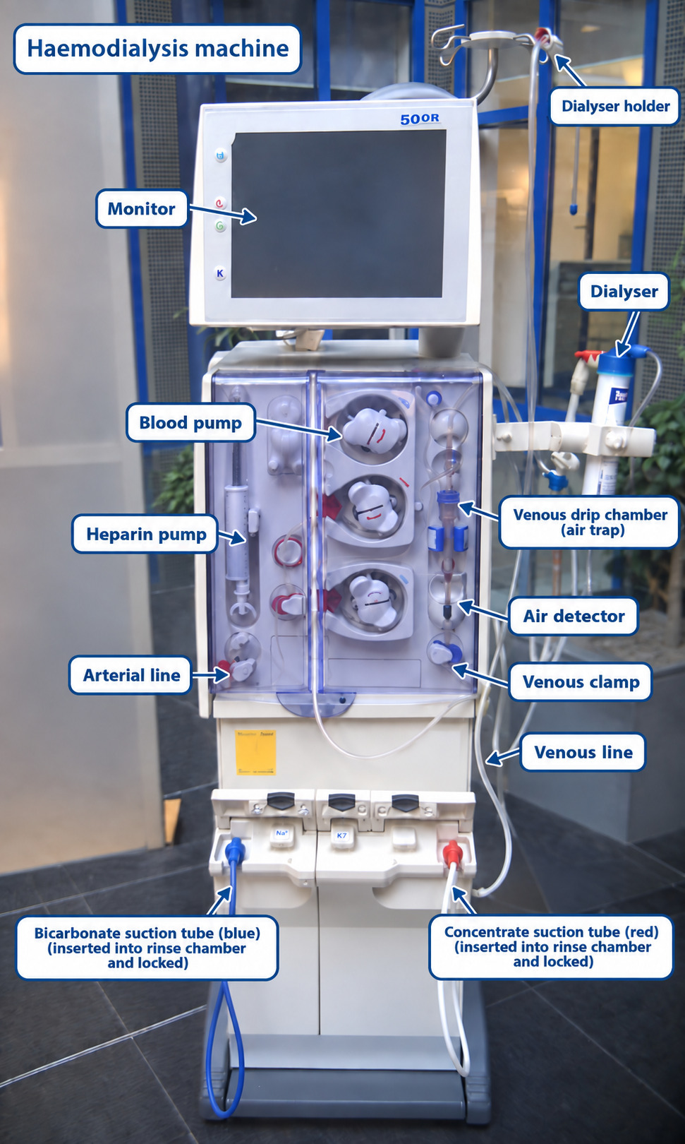
In principle, a haemodialysis machine consists of two connected systems: an extracorporeal blood circuit and a dialysate circuit. The blood circuit contains the blood pump, dialyser and blood tubing. The dialysate circuit prepares and delivers dialysate to the dialyser and removes the spent fluid. Both circuits are surrounded by monitoring, alarm and safety systems.
Modern machines increasingly combine measurements from the blood circuit, dialysate circuit and patient with software that displays treatment data in real time and records the course of each individual dialysis session.
Blood pump
The blood pump is usually a roller pump that compresses a short segment of silicone pump tubing. Typical prescribed blood-flow rates (Qb) are approximately 200–600 mL/min, depending on the vascular access, patient and treatment prescription.
If the selected pump speed exceeds the blood supply available through the arterial access needle or catheter lumen, the pressure measured before the pump becomes excessively negative. When the programmed alarm limit is crossed, the machine sounds an alarm and normally stops the blood pump. Treatment remains interrupted until the cause has been checked, the alarm cleared and the pump deliberately restarted.
Conversely, excessive resistance to blood returning to the patient raises the venous-line pressure. Crossing the upper venous-pressure limit likewise triggers an alarm and protective interruption.
In either situation the patient, vascular access and entire bloodline must be inspected. Possible causes include needle malposition, access stenosis, insufficient access flow, catheter malposition or thrombus, a kinked or compressed line, and patient movement. Pressure limits are patient- and access-specific; isolated pressure values should never replace clinical assessment.
Dialyser
The dialyser is the component in which solute and water exchange takes place. A rigid housing contains thousands of hollow fibres—by far the most common modern design—or, historically, parallel plates. Two ports carry blood and two carry dialysate, with the semipermeable membrane keeping the two fluid compartments separate.
The hollow fibres provide a large area for contact across the membrane. Commercial dialyser surface areas commonly range from about 0.5 to 2.2 m², although the appropriate size depends on the prescribed treatment.
Before connection to the patient, the extracorporeal circuit and dialyser are primed according to the manufacturer’s instructions to remove air and manufacturing or sterilisation residues. The dialyser’s blood-compartment volume is commonly in the approximate range of 40–150 mL, depending on the model.
Dialyser performance has two related but distinct dimensions: solute clearance and ultrafiltration. Clearance depends on membrane surface area and thickness, pore size and distribution, blood and dialysate flow, and the properties of the solute. “Efficiency” generally describes small-solute transfer and is often represented by the mass-transfer area coefficient, KoA. “Flux” describes hydraulic permeability and is represented by the ultrafiltration coefficient, KUF.
Ultrafiltration coefficient (KUF): the volume of water that can cross the membrane per unit time and transmembrane pressure, usually expressed in mL/h/mmHg. Values may range from only a few to more than 80 mL/h/mmHg. Labels such as low-, medium- and high-flux are not defined by one universal numerical threshold. High-permeability dialysers require precise volumetric ultrafiltration control.
Worked example:
A patient is 3 kg above target weight, and the prescribed treatment time is 4 hours. Ignoring any fluid given or consumed during treatment, the required net ultrafiltration rate is 3,000 ÷ 4 = 750 mL/h.
For a dialyser with a nominal KUF of 5 mL/h/mmHg, the simplified relation UFR = TMP × KUF gives a TMP of 150 mmHg.
This calculation is educational only. In clinical practice the machine controls net ultrafiltration volumetrically, and actual TMP reflects pressures throughout both blood and dialysate compartments, oncotic pressure, membrane fouling and changing treatment conditions. It is not safely derived from a single arterial-pressure reading.
Solute-transfer performance: urea KoA values supplied by manufacturers may span roughly 200–1,200 mL/min. Values below about 300 mL/min have traditionally been described as low efficiency and values above about 600 mL/min as high efficiency. Data sheets may also report clearances for creatinine, phosphate, vitamin B12 or inulin at specified blood and dialysate flows, as well as sieving coefficients.
The sieving coefficient describes how readily a substance is carried through a membrane by convection. It is usually defined as the solute concentration in the ultrafiltrate divided by the concentration in plasma water:
S = Cultrafiltrate / Cplasma water
A coefficient near 1 means the solute passes with water relatively freely; a coefficient near 0 means it is retained. An ideal dialysis membrane therefore has a coefficient close to 0 for albumin while permitting effective removal of the intended uraemic solutes.
Membrane cut-off and retention characteristics vary considerably. Conventional low-flux membranes mainly remove small solutes; high-flux and newer medium cut-off designs extend removal into the middle-molecule range while attempting to limit clinically important albumin loss. A single nominal “cut-off” does not fully describe a dialyser’s clinical performance.
Dialysers may be sterilised with steam, gamma irradiation, electron beam or ethylene oxide, depending on the product. Ethylene oxide is now uncommon for dialysers because residual exposure can cause hypersensitivity. Every dialyser and bloodline must be primed exactly as specified by its manufacturer.
Small solutes include electrolytes and urea. Middle and larger molecules include vitamin B12, β2-microglobulin, cystatin C and free light chains. Molecular mass is only one determinant of removal: protein binding, distribution volume, molecular shape, membrane adsorption and treatment modality also matter.
| Substance | Approximate molecular mass |
|---|---|
| Sodium (Na⁺) | 23 Da |
| Phosphorus (P) | 31 Da |
| Potassium (K⁺) | 39 Da |
| Urea | 60 Da |
| Phosphate | ≈95 Da |
| Creatinine | 113 Da |
| Uric acid | 168 Da |
| Glucose | 180 Da |
| Gentamicin | ≈478 Da |
| Vitamin B12 | 1,355 Da |
| Vancomycin | 1,449 Da |
| Inulin | ≈5,200 Da |
| β₂-microglobulin (β₂M) | 11,800 Da |
| Cystatin C | 13,300 Da |
| κ free light chain | ≈22,500 Da |
| λ free light chain | ≈45,000 Da |
| Albumin | ≈66,500 Da |
Dialysis membranes
Dialysis membranes have historically been made from unsubstituted cellulose, modified cellulose, mixed cellulosic-synthetic materials or fully synthetic polymers.
Unmodified cellulose membranes such as cellophane and cuprophan are now rarely used. Their exposed hydroxyl groups can activate complement and contribute to bio-incompatibility.
In modified or substituted cellulose membranes, chemical groups replace some of the hydroxyl groups of the original cellulose. Cellulosic-synthetic membranes, historically called hemophan or Cellosyn, add synthetic compounds to alter the blood-contact surface and improve biocompatibility.
Most contemporary dialysers use synthetic membranes. Common polymer families include polysulfone, polyethersulfone, polyacrylonitrile, polymethyl methacrylate and polyamide. These materials can be engineered for high hydraulic permeability and good mechanical strength.
Cellulose membranes tend to activate complement more strongly than modern synthetic membranes. Modified cellulose such as cellulose triacetate can nevertheless be biocompatible and is available in different permeability ranges. “Synthetic” does not mean that every membrane has identical adsorption, albumin loss or hypersensitivity characteristics; the exact product remains important.
High-flux synthetic membranes improve β2-microglobulin clearance compared with older low-flux membranes. Whether any one membrane material independently improves long-term survival is much less certain and should not be inferred solely from laboratory biocompatibility markers.
Some membranes have special surface treatments. Vitamin-E-coated membranes have been studied for oxidative-stress and biocompatibility effects, but evidence for major patient-centred benefits remains limited. The AN69 membrane has a negatively charged surface and is associated with bradykinin-mediated reactions in susceptible settings; the surface-treated AN69ST membrane adsorbs heparin and may support reduced-anticoagulation protocols in selected patients under a validated local protocol.
Membrane biocompatibility
A biomaterial can affect blood cells and plasma proteins as soon as blood contacts it. A comparatively biocompatible membrane provokes less complement and kallikrein–kinin activation, less cellular inflammatory activation, fewer undesirable protein interactions and less thrombogenic stimulation.
Biocompatibility depends on both membrane chemistry and dialyser design. Hydroxyl groups in unmodified cellulose can activate the alternative complement pathway and neutrophils. Substituted cellulose and synthetic membranes generally cause less complement activation, but individual synthetic products differ in protein adsorption and surface charge.
Potential features associated with better biocompatibility
Studies have examined whether more biocompatible or high-flux membranes are associated with:
less β2-microglobulin accumulation and dialysis-related amyloidosis;
fewer hypersensitivity reactions;
less intradialytic hypotension;
slower loss of residual kidney function after dialysis initiation;
lower infection or inflammation burden;
better nutritional or lipid markers; and
better long-term morbidity or survival.
The strength of evidence is not equal for all these outcomes. Current practice commonly favours biocompatible high-flux membranes, but membrane selection must also consider modality, albumin loss, allergy history, anticoagulation strategy, water quality, local resources and the individual prescription.
Hypersensitivity reactions during dialysis
“Hypersensitivity reaction during dialysis” is more accurate than assuming a membrane allergy. Potential triggers include the membrane or sterilant, dialysate contamination, anticoagulants, intravenous iron or other administered medicines, bradykinin generation, complement activation and other mechanisms.
These events have sometimes been called “first-use reactions”, but they can also occur with later exposures. Acute dyspnoea, wheeze, flushing, urticaria, angio-oedema, hypotension or collapse requires immediate interruption and emergency assessment according to the unit protocol. The extracorporeal blood should not automatically be returned when a severe reaction is suspected.
Blood tubing
The segment carrying blood from the patient to the dialyser is conventionally called the arterial line, and the segment returning blood from the dialyser to the patient is called the venous line.
Bloodline sets contain chambers in which air can separate from blood. Arterial pressure is usually measured before the pump, while venous pressure is measured after the dialyser in or near the venous chamber. The venous return path also passes an air detector. If air or an abnormally low chamber level is detected, the pump stops and a venous-line clamp closes to prevent air from reaching the patient.
Some systems add a post-pump arterial-pressure sensor. TMP is calculated from blood-side and dialysate-side pressure measurements; the exact formula depends on which pressures the machine measures.
Blood tubing may be sterilised with ethylene oxide or another validated method. The entire set must be inspected, assembled and primed according to the manufacturer’s instructions before patient connection.
Dialysate preparation and delivery
A machine normally mixes treated water with acid concentrate and bicarbonate concentrate for an individual station. Some facilities use central systems for part of the concentrate distribution, but final proportioning and safety checks occur at the dialysis machine.
The acid component is usually a liquid concentrate and may use acetate or citrate in a small amount as the acidifying agent. Bicarbonate may be supplied as liquid concentrate or prepared from dry powder or a cartridge.
Individual preparation provides prescription flexibility. Treated water is heated to the prescribed temperature, deaerated and proportioned with the concentrates. Dialysate is then pumped past the outside of the hollow fibres, normally counter-current to blood flow.
Dialysate delivery system
The delivery system must continuously produce dialysate of the prescribed composition, temperature and flow, deliver it to the dialyser, and supervise fluid removal and effluent disposal.
Its principal stages include:
Water supply and conditioning: appropriately treated dialysis water enters the machine, is heated—often within an adjustable range of about 34–39 °C—and is deaerated.
Proportioning: the machine mixes treated water with acid and bicarbonate concentrates in defined ratios to create dialysate with the intended electrolyte composition, pH, temperature and flow.
Independent safety checks: conductivity, temperature and sometimes pH sensors verify the fluid before it can reach the dialyser.
Balancing and ultrafiltration control: inflow and outflow are balanced so that net fluid removal matches the prescription.
The final dialysate composition depends on the compatible machine and concentrates selected by the dialysis service. Safe use requires correct concentrate connections, machine disinfection, water-quality surveillance, pre-treatment checks and an individual prescription at every session.
Electrical conductivity is used as a practical surrogate for total ionic concentration. Pure water conducts electricity poorly, whereas electrolyte solutions conduct it well. Conductivity therefore helps detect incorrect concentrate-to-water proportioning, but it does not identify every individual electrolyte concentration.
For a conventional sodium concentration near 135–140 mmol/L, conductivity is often around 13.5–14.5 mS/cm, depending on the complete formulation and temperature compensation. If conductivity or temperature is outside the programmed range, the machine places dialysate in bypass so it does not enter the dialyser and triggers an alarm. Independent control and monitoring sensors provide an additional layer of protection.
Ultrafiltration control
Modern machines use volumetric balancing chambers or accurate inlet/outlet flow measurement to control net ultrafiltration. Older pressure-control concepts adjusted dialysate pressure to obtain an estimated UF rate, but high-flux treatment requires volumetric control because membrane water permeability is high and changes during treatment.
The prescribed UF profile may be constant or deliberately varied during the session. Accuracy and permitted maximum rate are machine-specific and must be verified in the device documentation; neither a nominal percentage nor a maximum such as 4 L/h is universally applicable or necessarily safe for a particular patient.
Conductivity, temperature and pressure monitoring
Conductivity: continuous conductivity monitoring verifies proportioning. Some machines can deliberately vary dialysate sodium over time (“sodium profiling”). Any unexplained deviation can be dangerous; out-of-range dialysate is diverted to drain and treatment alarms.
Temperature: dialysate temperature is continuously monitored. A common clinical setting is close to 36–37 °C, with cooler dialysate sometimes prescribed to improve haemodynamic tolerance. Excessive heat can cause haemolysis and is a medical emergency. Values outside the machine’s validated safety limits trigger bypass and alarm.
Blood-leak detector: an optical detector monitors spent dialysate for blood, which can indicate a membrane rupture. Detection stops treatment flow or places the system in a safe state according to the machine design. Alarms must be investigated rather than simply overridden.
Arterial and venous pressure: sensors detect changes caused by access inflow problems, obstruction, kinking, clotting, needle position or disconnection. Alarm limits should be set around the patient’s expected pressures. Fixed statements such as “arterial above −100 mmHg” or “venous below 200 mmHg” are not universal safety limits.
Air detector: an ultrasonic sensor near the end of the venous line works with a bubble trap and automatic venous clamp. Detection of air stops the blood pump and clamps the return line.
Bubble trap
The venous chamber allows bubbles to rise out of the blood path. Together with the downstream air detector and venous clamp, it reduces the risk of air embolism. Correct blood level in the chamber and secure line connections remain essential.
Online treatment-dose monitoring
Some machines estimate the delivered dialysis dose during treatment. Ionic dialysance—derived from brief controlled changes in dialysate conductivity—is widely used as a surrogate for urea clearance. Other systems analyse ultraviolet absorbance in spent dialysate to follow removal of UV-absorbing solutes.
Combining estimated clearance with elapsed treatment time and an estimate of urea distribution volume provides an online Kt/V. This is useful for trend monitoring but does not always replace formal adequacy assessment with correctly timed blood samples. Machines may also measure dialysate outlet pressure for TMP calculations and include pH monitoring.
Patient monitoring systems
Blood pressure
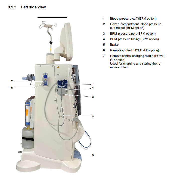
Many machines can automatically measure and record non-invasive blood pressure at programmed intervals. The reading must still be interpreted alongside symptoms, heart rate, UF rate and the patient’s baseline.
Electrocardiography
Selected systems or connected monitors can display heart rhythm, particularly in high-dependency or remotely supervised settings. This does not replace appropriate clinical monitoring when arrhythmia or ischaemia is suspected.
Relative blood-volume monitoring
Optical or ultrasonic sensors can track haematocrit or total protein concentration in the arterial bloodline. As plasma water is removed faster than vascular refilling replaces it, blood becomes concentrated; the device expresses this as a fall in relative blood volume (RBV).
RBV trends can be measured reproducibly within a session, and some devices use them in feedback-controlled UF or dialysate-sodium systems. A falling RBV may precede hypotension, but no single threshold reliably predicts symptoms in every patient.
Possible responses to developing haemodynamic instability include reassessing the patient, stopping or reducing UF, positioning, and administering fluid when clinically indicated. Increasing dialysate sodium or giving hypertonic fluid is not a routine automatic solution because it can increase thirst and interdialytic weight gain. Some monitors also estimate absolute haematocrit and oxygen saturation.
Fluid-status monitoring
Some blood-volume systems analyse the response to a brief UF change, and other technologies use bioimpedance or ultrasound. These measurements may support—but do not by themselves determine—target-weight assessment. Symptoms, blood pressure, examination, residual kidney function and longitudinal trends remain central.
Recirculation
Access recirculation can be estimated by thermodilution, saline dilution or conductivity methods. A machine may briefly alter dialysate temperature, or a defined saline bolus may be injected into the venous line while a sensor detects its appearance in the arterial line. Unexpected recirculation should prompt review of needle direction and spacing, line reversal, access flow and possible stenosis.
Delivered Kt/V
Delivered dose may be estimated online from ionic dialysance or spent-dialysate ultraviolet signals. The estimate is valuable for detecting an unexpectedly low clearance during the session, but laboratory urea modelling remains the reference method used by many programmes for periodic adequacy assessment.
Anticoagulant delivery
When systemic anticoagulation is appropriate, the machine’s syringe pump can deliver unfractionated heparin as a loading dose and/or infusion according to the prescription. Anticoagulation is not mandatory in every session: patients with active bleeding, recent surgery, heparin-induced thrombocytopenia or other contraindications require an alternative strategy and close circuit observation.
Sodium profiling
Some machines can vary dialysate sodium during the session by changing concentrate proportioning within validated limits. Although profiling can sometimes improve short-term haemodynamic tolerance, routine use may increase sodium balance, thirst and interdialytic weight gain. It should therefore be individualised and evaluated rather than used automatically.
Haemodiafiltration
Online haemodiafiltration combines diffusion with high-volume convection. A high-flux dialyser and controlled ultrafiltration increase middle-molecule removal, while sterile non-pyrogenic substitution fluid replaces the convective volume beyond the prescribed net fluid removal.
The technique requires precise volumetric control, ultrapure dialysate and online production of substitution fluid that meets pharmacopoeial sterility and endotoxin requirements. Modern machines produce it through a validated cascade of ultrafilters and continuously monitor the process. Recent randomised evidence supports a survival benefit from adequately dosed high-volume haemodiafiltration in eligible patients, but delivery depends on access flow, treatment time, haemoconcentration and local quality systems.
Single-needle and double-needle dialysis
Most machines support conventional double-needle dialysis, in which one needle continuously withdraws blood and the other continuously returns it. A single-needle mode can use one access needle connected through a Y-piece, alternating withdrawal and return phases rather than performing both simultaneously.
The advantage is a single puncture, which can be useful when cannulation options are limited. Its disadvantages are lower effective clearance, more complex cycling and a greater risk of recirculation.
A defined volume is drawn into the arterial circuit and then previously dialysed blood is returned. The method requires compatible bloodlines and a compliant expansion chamber or controlled pump cycle; some systems use two blood pumps. The prescription and machine setup must be performed by trained dialysis staff.
Selected evidence and technical standards
Key points
- The blood pump moves blood through the dialyser and back to the patient.
- The membrane separates blood from dialysate while allowing selective movement of water and solutes.
- Pressure sensors can help identify resistance, kinks, obstruction, or access problems.
- Air detectors, bubble traps, and pump-stop mechanisms are important safety layers.